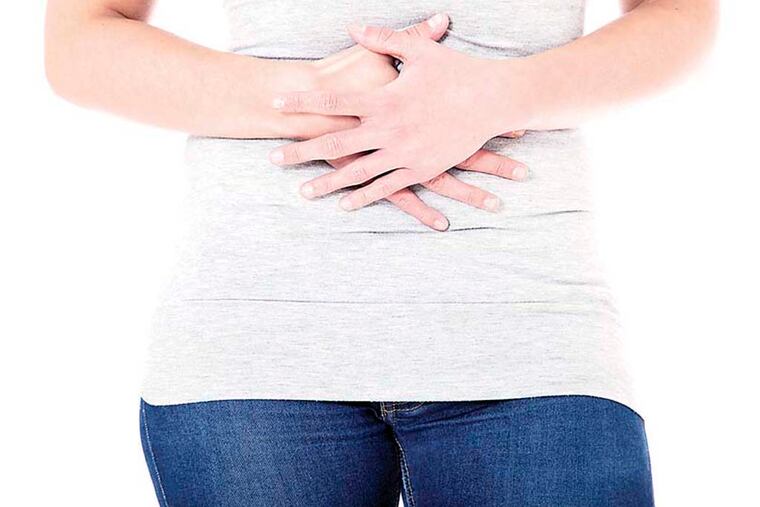
Abdominal pain continues during pregnancy
Early in her pregnancy with her first baby, the 31-year-old nurse felt nauseated and began having pain in her lower right abdomen. Her obstetrician explained that during the first trimester, it is quite common to feel sick to your stomach and, in all likelihood, the sharp twinges came from ligaments stretching to accommodate the growing fetus.

Early in her pregnancy with her first baby, the 31-year-old nurse felt nauseated and began having pain in her lower right abdomen. Her obstetrician explained that during the first trimester, it is quite common to feel sick to your stomach and, in all likelihood, the sharp twinges came from ligaments stretching to accommodate the growing fetus.
Weeks passed, and well into her second trimester, the woman still had trouble keeping food down. The pain on her lower right side subsided, but she flinched when anyone touched the upper right side of her belly.
She was thin from the start, only 120 pounds, and although her baby was growing normally, the woman steadily lost weight. Twice, tests found elevated white blood counts in her urine so she received antibiotics to treat the suspected infections.
Her weight continued to drop. Her doctors told her the problem was probably hyperemesis gravidarum, more commonly known as extreme morning sickness.
At least 60,000 women a year, and likely many more, suffer from the condition, which is believed to stem from rising hormone levels. Most women begin to feel better by the 14th week of pregnancy, but for about 20 percent, it lasts all the way through.
That appeared to be the case for this patient. A month before her due date, her weight had dropped to 100 pounds and she developed a high fever. Concerned that she might have chorioamnionitis, an infection in the amniotic fluid and the membranes surrounding the fetus, the woman was admitted to the hospital for an emergency C-section.
While her premature but otherwise healthy six-pound infant was cared for in the neonatal intensive care unit, the mother was examined by an infection specialist.
Laboratory results showed that she had a high white blood count in her urine, which might have indicated a kidney infection, but curiously, no bacteria were found in the sample.
Tests did identify E. coli in her bloodstream, however, and since she was still tender in the upper right portion of her abdomen, she was also checked for signs of liver problems and an ultrasound was done to examine her gallbladder.
Both organs appeared normal.
Solution
Her doctors thought that one possible source of the mother's illness might be hydronephrosis, a swelling of the kidney when urine backs up because it cannot drain from the bladder. The condition sometimes occurs near the end of pregnancy, when the baby grows so large that it presses on the mother's ureters.
They also ruled out chorioamnionitis because it almost always leads to endometritis, but the woman had no pain in her uterus.
A more in-depth study was made of her gallbladder, using a nuclear scan. If the gallbladder is healthy, radioactive dye in the organ will light up.
It lit up.
Stumped, the doctors sent the woman home with a prescription for oral antibiotics. She returned for a checkup several weeks later, looking and feeling well.
Although she still had not gained much weight, she was eating a little more than when she was pregnant. She still, however, had an unusually large number of white blood cells in her urine.
"If it is something bad," her doctor said, "it will declare itself." The mystery would not last forever.
One week later, the woman found herself back in the hospital with a fever and the same sharp pain in her lower right abdomen that she had felt during the first trimester.
A CAT scan revealed an abscess the size of a golf ball sitting on her bladder. Doctors drained the abscess and over three days, removed 50 ccs (about three tablespoons) of pus. Tests showed that it contained E. coli.
But no bacteria grew out of tissue removed from her bladder.
Could this be an infection in her ovary?
No. It was chronic appendicitis.
Early in her pregnancy, the woman developed appendicitis - causing the sharp pain in her lower right abdomen. The appendix probably ruptured at some point, but the infection was kept moderately under control by the antibiotics she received for the suspected urinary tract infection.
The reason that there were white cells in her urine but no bacteria made sense now. When an abscess develops outside the bladder, the bladder will produce white blood cells, but the bacteria will not be present.
As the baby grew and her uterus pushed her organs out of place, her cecum, the anatomical structure near the start of the large intestine, and appendix moved up into her abdominal cavity. That, her doctors surmised, was why the pain traveled to her upper right quadrant.
Their theory was supported by a study conducted in the 1930s, in which healthy women drank barium at various stages of their pregnancy and were X-rayed to follow the path of the cecum over nine months. (Yikes.)
This patient was given more antibiotics and asked to wait a few weeks before surgeons would decide whether to remove the remains of her appendix.
Unlike acute appendicitis, which normally requires immediate surgery, they explained, her chronic inflammation would make it too difficult to define the area that might need to be removed. Once antibiotics subdued the infection, a CAT scan would be done to determine if surgery was necessary.
After several days of intravenous treatment, the woman went home feeling well. She was steadily gaining weight and looking forward to her newborn's imminent release from the neonatal intensive care unit.